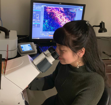
Over the next three years the Parkinson’s Foundation will invest more than $50 million to Parkinson’s disease (PD) research and clinical care. At the heart of our research initiatives are scientists and researchers who have received Foundation awards to improve our understanding of Parkinson’s, which will ultimately lead us to a cure.
Much of our understanding of Parkinson’s disease comes from genetic studies. The most common genetic changes linked to Parkinson’s occur in the GBA gene. The role of GBA in cells is to break down complex lipids in the cell’s “recycling bin,” the lysosome.
Roy N. Alcalay, MD, MS, is a Stanley Fahn Junior Faculty Awardee at Columbia University Medical Center who received a Parkinson’s Foundation grant to identify the parts of lipid metabolism that are most affected by Parkinson’s and to find potential drug targets to correct them. With cutting-edge technology, Dr. Alcalay’s research will measure 520 lipids in blood samples from people with Parkinson’s and from people without the disease. In people who carry Parkinson’s-related mutations in the GBA or alpha-synuclein genes, we will test if altered lipid levels are linked to Parkinson’s diagnosis.
Dr. Alcalay’s research will also analyze 600 people with Parkinson’s and 400 without the disease, testing for genetic changes in 32 genes involved with lipid metabolism. His team will look at the relationship between lipid levels and the activities of lysosome enzymes, also assessing genetic changes in the enzymes’ genes. Our hope is that Dr. Alcalay’s and his team can identify novel drug targets and biomarkers for Parkinson’s that will improve diagnosis and treatment of the disease.
The Parkinson's Foundation Stanley Fahn Junior Faculty Award helps ensure promising early career scientists stay in the PDresearchfield. This award provides junior investigators the support they need to develop their own independent funding source.
What's Next: Reporting Our Findings
Parkinson’s Foundation research awards fund Parkinson’s studies than can span up to three years. Scientists submit yearly progress reports to the Parkinson’s Foundation, and we report findings once the studies have concluded. Stay up to date with our latest research findings at Parkinson.org/Blog.
Protein May Hold Clues to Development of Parkinson’s
Over the next three years the Parkinson’s Foundation will invest more than $50 million to Parkinson’s disease (PD) research and clinical care. At the heart of our research initiatives are scientists and researchers who have received Foundation awards to improve our understanding of Parkinson’s, which will ultimately lead us to a cure.
Alpha-synuclein (αSyn)is a protein central to Parkinson’s. In Parkinson’s, this protein misfolds, forming a clump in the brain. Large clumps are known as “Lewy bodies” and disrupt the brain’s normal functioning in people with PD.
Alpha-synuclein is also involved in the regulation of lipids and fatty acids, which help to prevent disease-associated changes in the brain. In Parkinson’s, alpha-synuclein is destabilized. This makes the protein more likely to break down and clump together. What triggers the destabilization of alpha-synuclein in the human brain remains one of the most critical questions in the study of Parkinson’s.
Tim Bartels, MSc, PhD, from Brigham and Women's Hospital Inc., received a Parkinson’s Foundation research grant to gain a better understating of alpha-synuclein in Parkinson’s. He and his research team will analyze the interactions of lipids with different forms of alpha-synuclein in human brain samples. Dr. Bartels hopes to discover which lipids and fatty acids prevent alpha-synuclein aggregation and which ones promote aggregation.
He will also investigate the normal interaction of alpha-synuclein with fatty acids and lipids. Together, these approaches should suggest how to develop drugs that stabilize alpha-synuclein by mimicking the beneficial lipids and fatty acids.
Dr. Bartels team may also find a signature of specific lipids and fatty acids that are associated with PD. This could be an easily accessible biomarker — a biological molecule that is a sign of disease — for Parkinson’s. Having a biomarker for Parkinson’s could lead to earlier diagnosis of the disease. This can improve outcomes for people living with PD.
The Parkinson’s Foundation Stanley Fahn Junior Faculty Awardhelps ensure promising early career scientists stay in the PD research field. This award provides junior investigators the support they need to develop their own independent funding source.
What’s Next: Reporting Our Findings
Parkinson’s Foundation research awards fund Parkinson’s studies than can span up to three years. Scientists submit yearly progress reports to the Parkinson’s Foundation, and we report findings once the studies have concluded. Stay up to date with our latest research findings at Parkinson.org/Blog.
Simple Blood Draw Reveals Early Inflammation/Fatigue Connection
First described as a common symptom of Parkinson’s disease (PD) more than 20 years ago, PD-related fatigue remains an under-recognized, clinically significant, disabling symptom that can diminish quality of life (Herlofson et al., 2018; Kluger et al., 2016). Often occurring prior to movement symptoms of PD — tremor, rigidity and bradykinesia — (Chong, Albor, Wakade, & Morgan, 2018), PD-related fatigue doesn’t go away over time and getting more rest does not help (APDA, 2017).
The 2014 Parkinson’s Foundation Conference on Fatigue found that half of all people with PD reported fatigue as a major problem, and one-third shared that fatigue is their single most disabling symptom. The conference brought together a multidisciplinary group of experts, including a scientist studying fatigue in breast cancer. Research in breast cancer has shown that inflammation plays a large role in fatigue, even years after treatment when patients are in remission. One of the questions that came out of the 2014 conference was whether inflammation also plays a role in fatigue in PD.
Recently published in the journal, Acta Neurologica Scandinavica, a study titled, “Inflammation and fatigue in early, untreated Parkinson’s” (Herlofson et al., 2018) sought to explore the possible association of proinflammatory cytokines (a type of substance released by a cell that promotes inflammation) and fatigue in PD. Considered the boss of the immune system, cytokines are chemical messengers responsible for up-regulating (initiating), as well as down-regulating (turning off) the immune response. Studies have found that proinflammatory cytokines may play a role in PD and have been proposed to be part of an immune response to tissue damage (Williams-Gray et al., 2016)
Funded by the Parkinson’s Foundation, Karen Herlofson, MD, led a study measuring 13 different inflammatory markers and adhesion molecules (helps cells stick to one another), obtained with a simple blood-draw in people with PD who had also been assessed for fatigue levels.
The study recruited 212 participants who were newly diagnosed with PD and untreated. They were initially recruited from the Norwegian ParkWest Project — a population-based prospective longitudinal cohort study of newly diagnosed, untreated people with PD. However, after excluding those with decreased cognitive function, symptoms of depression, excessive daytime sleepiness, apathy, other diseases or relevant medications, a total of 47 participated in this study; 24 had low fatigue scores and 23 had high fatigue scores.
Demographic data (age, years of education, weight, and height) were collected during a semi-structured interview. Disease severity was assessed by the Unified Parkinson’s Disease rating Scale part III (UPDRS III). Fatigue was assessed by the Fatigue Severity Scale (FSS), a self-administered questionnaire, which focuses upon the physical social, and mental aspects of fatigue. All 47 participants had their blood drawn the same day as the clinical assessment and all fasted the night before. The blood draw occurred between 8 and 10 a.m., to minimize confounding factors caused by circadian rhythm.
Results
Compared to the study participants without fatigue, participants with fatigue:
Had significantly higher levels of the cytokine IL1-Ra and the adhesion molecule VCAM-1
Had more advanced disease, as measured by the Unified Parkinson’s Disease Rating Scale (UPDRS) motor score.
Had lower cognitive function, as measured by the MMSE (Mini-Mental State Examination).
Had more depression, as measured by the Montgomery-Åsberg Depression Rating Scale (MADRS).
Had less excessive daytime sleepiness, as measured by the Epworth Sleepiness Scale (ESS), showed no correlations between IL1-Ra or VCAM-1 with age, sex, years of education, BMI, UPDRS, MADRS, MMSE, apathy, or ESS.
What Does This Mean?
PD-associated fatigue can have a detrimental impact on quality of life; yet, the underlying biological cause remains unknown. This study found that having higher blood levels of the inflammatory markers IL1-Ra and VCAM-1 were associated with higher fatigue levels in newly diagnosed, untreated participants with PD. If true, and an altered immune response is indeed a factor, this finding may offer new targets to explore for future treatment — as the immune system is a promising therapeutic target for disease modification.
Additionally, these findings may, if correct, offer some future early PD diagnostic potential — as fatigue commonly presents before the classic hallmark symptoms of PD. We know that inflammation contributes to neurodegeneration in the brain. Whether or not cytokines are increased as a result of the stress and tissue damage that are a consequence of PD — or whether the inflammation happens prior to the fatigue — is a question that must be answered. This study brings us one step closer
Learn More
The Parkinson’s Foundation believes in empowering the Parkinson’s community through education. Learn more about the connection between fatigue and Parkinson’s in the below Parkinson’s Foundation resources or by calling our free Helpline at 1-800-4PD-INFO (473-4636).
Chong, R., Albor, L., Wakade, C., & Morgan, J. (2018). The dimensionality of fatigue in Parkinson's disease. J Transl Med, 16(1), 192. doi:10.1186/s12967-018-1554-z
Herlofson, K., Heijnen, C. J., Lange, J., Alves, G., Tysnes, O. B., Friedman, J. H., & Fagundes, C. P. (2018). Inflammation and fatigue in early, untreated Parkinson's Disease. Acta Neurol Scand, 138(5), 394-399. doi:10.1111/ane.12977
Kluger, B. M., Herlofson, K., Chou, K. L., Lou, J. S., Goetz, C. G., Lang, A. E., Friedman, J. (2016). Parkinson's disease-related fatigue: A case definition and recommendations for clinical research. Mov Disord, 31(5), 625-631. doi:10.1002/mds.26511
Williams-Gray, C. H., Wijeyekoon, R., Yarnall, A. J., Lawson, R. A., Breen, D. P., Evans, J. R., group, I.-P. s. (2016). Serum immune markers and disease progression in an incident Parkinson's disease cohort (ICICLE-PD). Mov Disord, 31(7), 995-1003. doi:10.1002/mds.26563
Gene Mutation That Promotes Protein Aggregation Could Spur Parkinson's
Over the next three years the Parkinson’s Foundation will invest more than $50 million to Parkinson’s disease (PD) research and clinical care. At the heart of our research initiatives are scientists and researchers who have received Foundation awards to improve our understanding of Parkinson’s, which will ultimately lead us to a cure.
A key feature of Parkinson’s disease is abnormal protein clumping within nerve cells or neurons. These protein clumps, called aggregates, spread throughout the nervous system as Parkinson’s progresses. How this occurs remains unclear. Mutations in the GBA1 gene are a strong genetic risk factor for developing Parkinson’s. They are also linked with faster progression of motor and cognitive symptoms.
Marie Ynez Davis, MD, PhD, is a Parkinson’s Foundation Clinical Research Awardee of VA Puget Sound, is investigating a potential new role for the gene GBA1 in speeding the spread of protein aggregates through exosomes. These are small bubble-like structures released by neurons and other cells. They contain proteins and other material that can travel and be received by other neurons and cells.
Dr. Davis’ goal is to find out whether a lack of GBA1 influences the development of Parkinson’s by increasing the number of exosomes and the proteins inside them that can be delivered to other neurons. This could promote clumping in the receiving neurons throughout the nervous system.
To achieve this goal, she will study human neurons from people with GBA1 mutations and Parkinson’s. She will examine the development and structure of exosomes. She will also look at their ability to promote protein aggregation.
Our hope is that results from this work will improve our understanding of how Parkinson’s occurs. It may also reveal new targets for therapies that could halt or slow progression of the disease.
Parkinson’s Foundation Clinical Research Awards help facilitate the development of clinician scientists, ensuring that promising early career scientists stay in the PD researchto help us solve, treat and end this disease. These scientists are vital, as they apply research to the real-world as well as applying real-world issues to research.
What’s Next: Reporting Our Findings
Parkinson’s Foundation research awards fund Parkinson’s studies than can span up to three years. Scientists submit yearly progress reports to the Parkinson’s Foundation, and we report findings once the studies have concluded. Stay up to date with our latest research findings at Parkinson.org/Blog.
Levodopa-Induced Dyskinesias Reversed in Gene Therapy Study
Levodopa-induced dyskinesias (LIDs) are a debilitating side effect of long-term use of levodopa therapy that negatively impacts the quality of life for upwards of 90% of people with Parkinson’s disease (PD). From the Greek word, dys, meaning ill or bad, and kīnēsia, meaning movement or muscular activity, dyskinesias are abnormal, involuntary muscle movements, often presenting as erratic writhing movements of the face, arms, legs or trunk.
This side effect is caused by the loss of dopamine in the striatum (a structure in the brain). That loss of dopamine causes the calcium channels in the brain to not function properly.
Why do calcium channels matter?
The human brain has roughly 100 billion interconnected cells. A calcium channel lets calcium into each cell, allowing it to function normally. Previous studies in rats have shown that calcium activity in the brain plays an important role in the onset of Parkinson’s. These channels are also believed to contribute to levodopa-induced dyskinesias.
Evidence suggests that silencing one specific type of these calcium channels might hold some therapeutic promise. The Cav1.3 channel is the calcium channel found in the striatum and is made by the human CACNA1D gene. There are existing drugs that can inhibit these calcium channels. However, these drugs are not very potent. For people with PD, they only partially help with levodopa-induced dyskinesias and the relief only lasts a short time, but hope may be on the horizon.
Gene Expression and Gene Silencing
Genes tell cells how to make proteins. This instruction process is called gene expression. Like a dimmer switch, gene silencing is the regulation of gene expression in a cell that prevents or reduces that expression. Figuring out how to identify and silence certain genes is being increasingly studied to help find new therapeutic avenues to combat diseases.
Funded by the Parkinson’s Foundation and the National Institute of Neurological Disorders and Stroke, the study “Genetic Silencing of Striatal CaV1.3 Prevents and Ameliorates Levodopa Dyskinesia” (Steece-Collier et al., 2019) investigated whether silencing of striatal CaV1.3 channels genetically ― as opposed to using medications ― might have the potential to transform Parkinson’s treatments. If successful, this approach could lead to the development of new highly targeted drugs that would allow people with PD to enjoy the motor benefits of levodopa, without the debilitating LID side effects.
To validate the CaV1.3 as a potential target for drugs that might reduce the LID, Kathy Steece-Collier, PhD, and her research team created a virus in their lab that delivers genetic material into cells to target the CaV1.3 channel and prevent it from being made. This virus is a stripped-down version of a normal virus, with all the harmful viral information removed, and is called a viral vector. They injected the viral vector into severely parkinsonian rats to see if that would reduce expression of CaV1.3 channels and thereby impact LIDs.
This was a two-month study that included a control group of severely parkinsonian rats for comparison (that would not receive the genetic treatment). Both groups of rats received increasing doses of Levodopa over time. All rats were regularly tested both prior to and after introducing levodopa therapy.
Results
If the CaV1.3 channels are silenced in severely parkinsonian rats prior to giving them levodopa therapy, this was able to completely prevent LID symptoms from forming.
Even with very high doses of daily levodopa, the prevention of LIDs remained and did so, long-term.
This experimental approach was capable of even partially reversing the LID in rats with established, preexisting severe LID behavior. Current calcium channel drugs are not able to do this.
The positive therapeutic (motor) benefits of levodopa remained throughout gene silencing.
As expected, in group of rats who did not receive the genetic treatment, LID worsened severely as the levodopa dose was increased.
What Does This Mean?
This study suggests that reducing the number of CaV1.3 calcium channels has the potential to transform the treatment of individuals with PD by allowing maintenance of the motor benefit of levodopa without the debilitating side effect of LIDs. Further, this study demonstrated it was possible to reverse preexisting, severe levodopa-induced dyskinesias. So, not only did the experimental approach have a protective/preventative effect, it helped the rats that were already severely impaired.
What we don’t know, yet, is whether or not reducing striatal CaV1.3 expression will work in people. However, the authors of this article stated that if the findings of this study can be validated clinically in people, this would be “a much-needed breakthrough in the treatment of individuals with PD and has the potential to allow the most powerful antiparkinsonian therapy identified (i.e., levodopa) to work unabated through the duration of the disease.”
In short, though there is much more work that lies ahead, this preclinical data suggests it may indeed be possible to prevent, and even reverse LIDs, which would be an extraordinary finding for the PD community.
Learn More
The Parkinson’s Foundation believes in empowering the Parkinson’s community through education. Learn more about the Parkinson’s and LID in the below Parkinson’s Foundation resources or by calling our free Helpline at 1-800-4PD-INFO (473-4636).
Chan, C. S., Guzman, J. N., Ilijic, E., Mercer, J. N., Rick, C., Tkatch, T., . . . Surmeier, D. J. (2007). 'Rejuvenation' protects neurons in mouse models of Parkinson's disease. Nature, 447(7148), 1081-1086. doi:10.1038/nature05865
Ovallath, S., & Sulthana, B. (2017). Levodopa: History and Therapeutic Applications. Ann Indian Acad Neurol, 20(3), 185-189. doi:10.4103/aian.AIAN_241_17
Steece-Collier, K., Stancati, J. A., Collier, N. J., Sandoval, I. M., Mercado, N. M., Sortwell, C. E., . . . Manfredsson, F. P. (2019). Genetic silencing of striatal CaV1.3 prevents and ameliorates levodopa dyskinesia. Mov Disord, 34(5), 697-707. doi:10.1002/mds.27695
Parkinson’s disease (PD) results in the loss of dopamine-producing cells in the brain. Because dopamine cannot cross the blood-brain barrier (a safety feature of the brain), people cannot simply take dopamine pills. The drug levodopa (L-dopa) — a precursor to dopamine and the gold standard for treating PD — does have the ability to cross the blood-brain barrier, where it successfully converts into the much-needed dopamine.
Since 1971, scientists were aware that one or more of the many microorganisms living in the human gastrointestinal tract prematurely convert the L-dopa into dopamine before it gets into the brain― but they didn’t know which ones. To combat some of this loss, L-dopa is usually combined with carbidopa to help block the premature metabolism, as well as help with the common side effects of nausea and vomiting.
Only about 44% of the L-dopa makes it to the brain. There are many different types of microorganisms, such as bacteria, viruses and fungi. Understanding which ones may affect this process could help improve the efficiency of levodopa as a treatment for PD.
A study titled, “Discovery and inhibition of an interspecies gut bacterial pathway for Levodopa metabolism” (Maini Rekdal, Bess, Bisanz, Turnbaugh, & Balskus, 2019), sought to decipher the molecular mechanisms by which gut bacteria might be responsible for this degradation of L-dopa en route to the brain. A complex undertaking, researchers used several, highly sophisticated methods, from running tests on the microbiome (trillions of microorganisms that help keep the body running smoothly) to evaluating inhibitors and testing the gut microbiota of people with and without PD.
Results
In the gut, some of the L-dopa was converted into dopamine by an enzyme from the common gut bacteria (Enterococcus faecalis).
Then, the dopamine (still in the gut) was further metabolized into a compound called m-tyramine, by an enzyme from a second bacterium (called Eggerthella lenta).
The enzymes from these two bacteria (Enterococcus faecalis and Eggerthella lenta) appear to be responsible for L-dopa metabolizing first into dopamine and then into m- tyramine, prior to reaching to blood-brain barrier, thereby diminishing how much L-dopa and the dopamine treatment successfully makes it to the brain.
Carbidopa is successful in inhibiting the metabolism of L-dopa by human enzymes, but not by the bacterial enzymes in the human gut.
When L-dopa was given to lab rodents orally along with a bacterial enzyme inhibitor called AFMT, this prevented metabolism of L-dopa and resulted in improved bioavailability of the drug in the animals’ blood stream.
What Does This Mean?
Using a complex set of steps, this study identified two enzymes in human gut microbiota (called Enterococcus faecalis and Eggerthella lenta) that are key to impeding a significant portion of the L-dopa from reaching the brain.
Further, in lab rodents, researchers increased the amount of L-dopa reaching the brain, by manipulating the activity of these enzymes in the gut with a drug, AFMT. This suggests that this approach (inhibiting bacterial enzymes) could possibly be developed for human use. In short, they may have found a drug that can reduce the premature metabolism of L-dopa into dopamine in the human gut, ultimately improving the efficiency of L-dopa treatment.
Maini Rekdal, V., Bess, E. N., Bisanz, J. E., Turnbaugh, P. J., & Balskus, E. P. (2019). Discovery and inhibition of an interspecies gut bacterial pathway for Levodopa metabolism. Science, 364(6445). doi:10.1126/science.aau6323
Vitamin K2 in People with Parkinson's with Mitochondrial Mutation
Over the next three years the Parkinson’s Foundation will invest more than $50 million to Parkinson’s disease (PD) research and clinical care. At the heart of our research initiatives are scientists and researchers who have received Foundation awards to improve our understanding of Parkinson’s, which will ultimately lead us to a cure.
Many cellular components are involved in the development and progression of Parkinson’s disease. One component is the “energy factory” of the cell, called mitochondria. Certain genetic changes are known to impair the function of mitochondria.
Jannik Prasuhn, MD, of the Institute of Neurogenetics, University of Luebeck in Germany, has received a Clinical Research Award from the Parkinson’s Foundation to investigate the use of vitamin K2 in people with Parkinson’s whose disease is caused by genetic mutations that affect the mitochondria.
Vitamin K2 has been shown to be safe in previous studies of people with PD with osteoporosis. The vitamin is known to play a role in supplying cells with energy.
Dr. Prasuhn’s goal is to determine if vitamin K2 can increase energy levels and improve symptoms in people with mitochondrial-related Parkinson’s.
To achieve this goal, he will study 130 people: 26 people with PD who have mitochondrial mutations; 52 people with PD with idiopathic PD, and 52 people without Parkinson’s. They will be randomly assigned to receive vitamin K2 or a placebo for six months. All participants will undergo brain imaging before and after the study, and they will undergo medical exams five times throughout the study.
If this research suggests K2 could be helpful to people with Parkinson’s who have mitochondrial mutations, our hope is that larger, longer-term studies will be conducted that could ultimately lead to using K2 as a PD treatment.
Parkinson’s Foundation Clinical Research Awards help facilitate the development of clinician scientists, ensuring that promising early career scientists stay in the PD researchto help us solve, treat and end this disease. These scientists are vital, as they apply research to the real-world as well as applying real-world issues to research.
What’s Next: Reporting Our Findings
Parkinson’s Foundation research awards fund Parkinson’s studies than can span up to three years. We report findings once the studies have concluded. Stay up to date with our latest research findings at Parkinson.org/Blog.
Ask the Experts: The Challenges of Using Marijuana as a Parkinson's Treatment, Part 2
Medical marijuana, or cannabis, is one of the most popular topics among the Parkinson’s disease (PD) community ― for people with PD, health professionals and researchers, alike. Earlier this year, the Parkinson’s Foundation hosted its first-ever convening on marijuana and Parkinson’s. Among the 46 attendees, of which 17 gave presentations, there was a reoccurring theme: what are the biggest hurdles the PD community faces when it comes to medical marijuana?
This is the second article in a two-part series. Read part one here.
Adverse Effects
Some of the most common side effects of cannabis-based products include:
drowsiness and fatigue
dizziness
dry mouth
anxiety
nausea
cognitive effects
Specifically, for smoked forms, side effects include cough, increased phlegm and bronchitis. Some rare but important side effects to note include: orthostatic hypotension, paranoia, depression, worsening of coordination of movement and rapid beating of the heart.
Specifically, regarding cognitive function, one review article of several studies found that attention and concentration were impaired in the short term (0-6 hours after use) but largely returned to normal in the longer term (three weeks or longer after use). However, decision making and risk taking were impaired three weeks or more after last cannabis use. Working memory was impaired shortly after use, but did not see any residual or long-term effects. Mixed results were seen as to whether there are long-term effects on impulsivity and verbal fluency after cannabis use.
→ Danny Bega, MD, MSCI, from Northwestern University Feinberg School of Medicine, spoke about this topic at the marijuana convening.
Finding the Right Formula
Physicians and pharmacologists are constantly trying to define the limits of their practice when it comes to cannabis. Not only must researchers find the right formula, they must also find the right delivery method. Cannabis can be delivered in various forms, from liquids to e-liquid (vapor) and inhalers to patches.
One additional challenge is that cannabis products are not highly regulated, so there can be a lot of variation from product to product and even from batch to batch within a single product. This needs to be regulated more so that people can know what is in the product they are purchasing and trust that it is safe.
→ Bill Arnold, CEO of Cannoid, LLC, spoke about this topic at the marijuana convening.
The Effects of Cannabis and Pain on Men VS Women
Of the 20 common conditions that qualify for medical marijuana, chronic pain has substantial evidence supporting the use of cannabis. People with PD report pain as one of the most common non-motor symptoms, which is not always responsive to pain medications.
PD-related pain is most common among women. Differences between men and women specifically in their susceptibility to intoxication and abuse liability have not been studied. Preclinical evidence suggests that female laboratory animals are more sensitive to cannabinoid (THC) relative to males in terms of treating pain, but they are also more sensitive to the abuse-related effects of these drugs. However, female animals develop tolerance to the pain-relieving effects of THC at a faster rate than males, rendering THC close to ineffective in females.
Ziva D. Cooper, PhD, and her colleagues tested cannabis to see if these findings in animals would translate to humans. Her study found that women who heavily smoke cannabis did not show a pain-relieving response, whereas men did. Regardless of pain response, women reported feeling as intoxicated as men and reported liking the cannabis as much as men.
Future studies investigating the use of cannabis and cannabinoids for PD-related pain are warranted. These studies should consider differences between men and women, cannabis experience and adverse effects.
→ Ziva D. Cooper, PhD, from the UCLA Cannabis Research Initiative, spoke about this topic at the marijuana convening.
The medical marijuana convening brought together a diverse group of experts from academia, clinics, industry, government and the Parkinson's community to establish a consensus on medical marijuana use in PD. The Parkinson’s Foundation will publish its findings on the convening in summer 2020.
Ask the Experts: The Challenges of Using Marijuana as a Parkinson’s Treatment, Part 1
Medical marijuana, or cannabis, is one of the most popular topics among the Parkinson’s disease (PD) community ― for people with PD, health professionals and researchers, alike. Earlier this year, the Parkinson’s Foundation hosted its first-ever convening on marijuana and Parkinson’s. Among the 46 attendees, of which 17 gave presentations, there was a reoccurring theme: what are the biggest hurdles the PD community faces when it comes to medical marijuana?
This is the first article in a two-part series. Read part two here.
Treating Parkinson’s Symptoms with Cannabis
There is not enough evidence yet to support that medical marijuana can help manage Parkinson’s symptoms, however there are studies on the topic. Unfortunately, they have mixed results. Generally, the studies have been small and some with no control groups. The effects of medical marijuana are not completely understood, especially in the PD population. The bottom line is that more studies are needed, specifically larger and more rigorously conducted studies.
Based on some observational studies, cannabinoids (the active molecules in marijuana) may potentially benefit some non-motor symptoms of PD including pain, anxiety, sleep problems (insomnia, RBD, RLS), weight loss and nausea. Potential adverse effects include dizziness, blurring of vision, loss of balance, mood and behavioral changes, hallucinations, and impaired cognition and motivation. Better studies are necessary to confirm these benefit and adverse effects for people with PD.
Controlled clinical trials of cannabinoids (where some people receive the drug and some do not) have reported mixed results for treating motor symptoms and levodopa-induced dyskinesia as well as improving quality of life.
While stories and videos exist showing that marijuana can treat PD symptoms, the challenge is showing that cannabis is better and safer than treatments that are currently available. A recent survey shows that the health community does not have a consensus on using cannabis as a treatment. This reflects lack of data, knowledge and training on the subject.
Future studies about medical marijuana and Parkinson’s should follow the highest standards of clinical trials to focus on:
Delivery type: do specific strains, soft gels, tinctures (alcohol-based cannabis extract), e-liquid (vapor), topicals, infused food, flower products, inhalers and patches treat symptoms differently and have different side effect profiles?
Dosage: what is the minimum dosage to guarantee effectiveness, what is the maximum dose tolerated and what dose will have a sustained benefit? Furthermore, how does this differ by strain and formulation?
Effect on motor vs non-motor symptoms: which symptoms can improve, worsen or stay the same with cannabis use?
Interaction with PD medications: how does cannabis interact with medications taken for PD symptoms?
Key component: What components of cannabis/marijuana provide the best response in PD with the least risk of side effects? What is the optimal CBD (Cannabidiol) to THC ratio?
PD-specific side effects: are people with PD uniquely susceptible to certain side effects that are not seen in the general population?
Lastly, there needs to be a widespread physician education on using cannabis as a treatment ― almost all physicians surveyed agreed that medical school curriculums should include education on cannabis.
→ Danny Bega, MD, MSCI, from Northwestern University Feinberg School of Medicine; Joseph Jankovic, MD, from Baylor College of Medicine; and Karl Kieburtz, MD, MPH, from the University of Rochester, spoke about this topic at the marijuana convening.
Potential Drug Interactions
One surprising fact shared at the meeting is that cannabis-based products have the potential to interact with other medications. Given that people with Parkinson’s may be on multiple medications for other conditions, it is important to be aware of these interactions to avoid complications.
Epidiolex® is the first FDA-approved cannabinoid prescription drug. It is an oral solution of cannabidiol most commonly used to treat rare forms of epilepsy. It has been shown to have interactions with many anti-seizure medications, some antibiotics and medications for lowering cholesterol, pain, anxiety, depression and blood pressure. In some cases, Epidiolex can make these medications more or less potent. In other cases, these medications can make Epidiolex more or less potent. Because Epidiolex largely contains cannabidiol, there is the possibility that other cannabis-based products may also interact with medications in a similar way.
Delta-9-tetrahydrocannibinol (THC) is the primary psychoactive component of marijuana (the part that gives a “high”). It can take a long time to take effect and cannot be easily measured for a therapeutic or medicinal dose. THC can also interact with certain medications such as valproic acid (for bipolar disorder, seizures, and migraines) and can result in increased psychoactive effects of marijuana.
Medical marijuana can be taken in an edible form. Care should be taken with this form, as it takes longer to feel an effect and lasts longer (4-8 hours as opposed to 2-3 hours for smoking or vaporizing). Often, because the effects are slow, people increase their dose, eating more, which can be dangerous. Edibles may also have more toxicity than smoked marijuana, because they are broken down by the liver into more toxic chemicals.
→ Jacqueline Bainbridge, PharmD, FCCP, MSCS, from the University of Colorado, spoke about this topic at the marijuana convening.
The medical marijuana convening brought together a diverse group of experts from academia, clinics, industry, government and the Parkinson's community to establish a consensus on medical marijuana use in PD. The Parkinson’s Foundation will publish its findings on the convening in summer 2020.
One of the most common genetic risk factors for Parkinson’s disease (PD) is having a mutated GBA gene (which makes the enzyme glucocerebrosidase). In fact, 5 to 10 percent of people with PD have that specific GBA mutation in one copy of the gene (mutations in both copies of the gene lead to Gaucher disease). It is more common than other genetic mutations associated with PD such as LRKK2, α-synuclein (SNCA) and PARK2. People with PD who have the GBA mutation tend to experience motor symptom deficits sooner, cognitive decline more rapidly and have particular difficulty with their gait (walking) and postural balance.
When GBA works as it should, it helps code (provide instructions) for making a digestive enzyme that breaks down potentially harmful substances, getting rid of unwanted bacteria and performs various housekeeping duties, including recycling worn out cell components. If both copies of the GBA gene are damaged (mutated), this negatively impacts a cascade of essential processes — which can lead to a dangerous build-up of toxins that harm the spleen, liver, lungs, bone marrow and brain (Gaucher disease). To date, most studies assessing GBA-related PD risk have been performed primarily in European and Asian-derived populations, while very few studies have been done in other populations, such as in the Latino population of South America.
A recently published study, titled, “The distribution and risk effect of GBA variants in a large cohort of PD from Columbia and Peru” (Velez-Pardo et al., 2019) sought to remedy that shortfall and characterize the frequency and distribution of GBA variants (variations in the gene). Specifically, Valez-Pardo et al. (2019) sequenced the entire GBA coding region in 602 participants with PD and 319 controls (people who do not have PD) from Colombia and Peru — all of whom were enrolled in the Latin American Research Consortium on the Genetics of Parkinson's disease. Age at enrollment was comparable for all participants. All study participants were evaluated by a movement disorder specialist at each site and met UK PD Society Brain Bank criteria for PD. The study used a blood test to sequence DNA from each participant.
Results
A significantly higher proportion of GBA mutation carriers (mutation in one copy of the gene) were seen in people with PD compared to people without Parkinson’s.
Those with GBA mutations from Peru and Columbia have a 4- and 6-fold increase in PD risk, respectively.
The age at onset was significantly earlier in GBA carriers (about 8 years) when compared to people with PD who were non-carriers.
A novel population-specific GBA mutation called, p.K198E was found only in Colombian participants. This was the first study to identify the p.K198E mutation.
Frequency of GBA mutations in Colombian participants was more than double most populations, e.g., Peruvian participants and European-derived populations. Note: This was primarily due to the presence of a population-specific mutation, p.K198E.
What Does It Mean?
While the specific biological mechanisms are still unclear, this study found that genetic changes in the GBA gene is linked to a higher risk of developing PD in both Columbians and Peruvians. Further, the novel mutation (p.K198E) the authors identified, found exclusively in the Columbian population, suggests there may be more yet to be discovered mutations in Latin American populations, that could provide unique insights into the disease mechanism.
Moreover, as nearly 10% of the Columbian population carries the GBA mutation — as opposed to about only 4% in other populations — the Columbian PD population may be particularly well suited as a group to study novel therapeutic approaches targeting GBA-related PD.
Learn More
The Parkinson’s Foundation believes in empowering the Parkinson’s community through education. Learn more about GBA and common PD mutations by vising the below Parkinson’s Foundation resources or by calling our free Helpline at 1-800-4PD-INFO (473-4636) for answers to all your Parkinson’s questions.
Sidransky, E., & Lopez, G. (2012). The link between the GBA gene and parkinsonism. Lancet Neurol, 11(11), 986-998. doi:10.1016/S1474-4422(12)70190-4
Velez-Pardo, C., Lorenzo-Betancor, O., Jimenez-Del-Rio, M., Moreno, S., Lopera, F., Cornejo-Olivas, M., . . . Mata, I. F. (2019). The distribution and risk effect of GBA variants in a large cohort of PD patients from Colombia and Peru. Parkinsonism Relat Disord. doi:10.1016/j.parkreldis.2019.01.030